
We do not diagnose disease or recommend a treatment protocol or dietary supplement for the treatment of disease. You should share this information with your physician who can determine what nutrition and disease treatment regimen is best for you. Ask your physician any questions you have concerning your medical condition.
You can search this site or the web for topics of interest that I may have written (use Dr Simone and topic). “We provide truthful information without emotion or influence from the medical establishment, pharmaceutical industry, national organizations, special interest groups or government agencies.” Charles B Simone, M.MS., M.D.
PATHWAYS TO THROMBOSIS DURING INITIAL AND RECURRENT INFECTIONS LIKE COVID AND THERAPEUTIC PLASMA EXCHANGE (TPE)
https://tinyurl.com/y48nu767
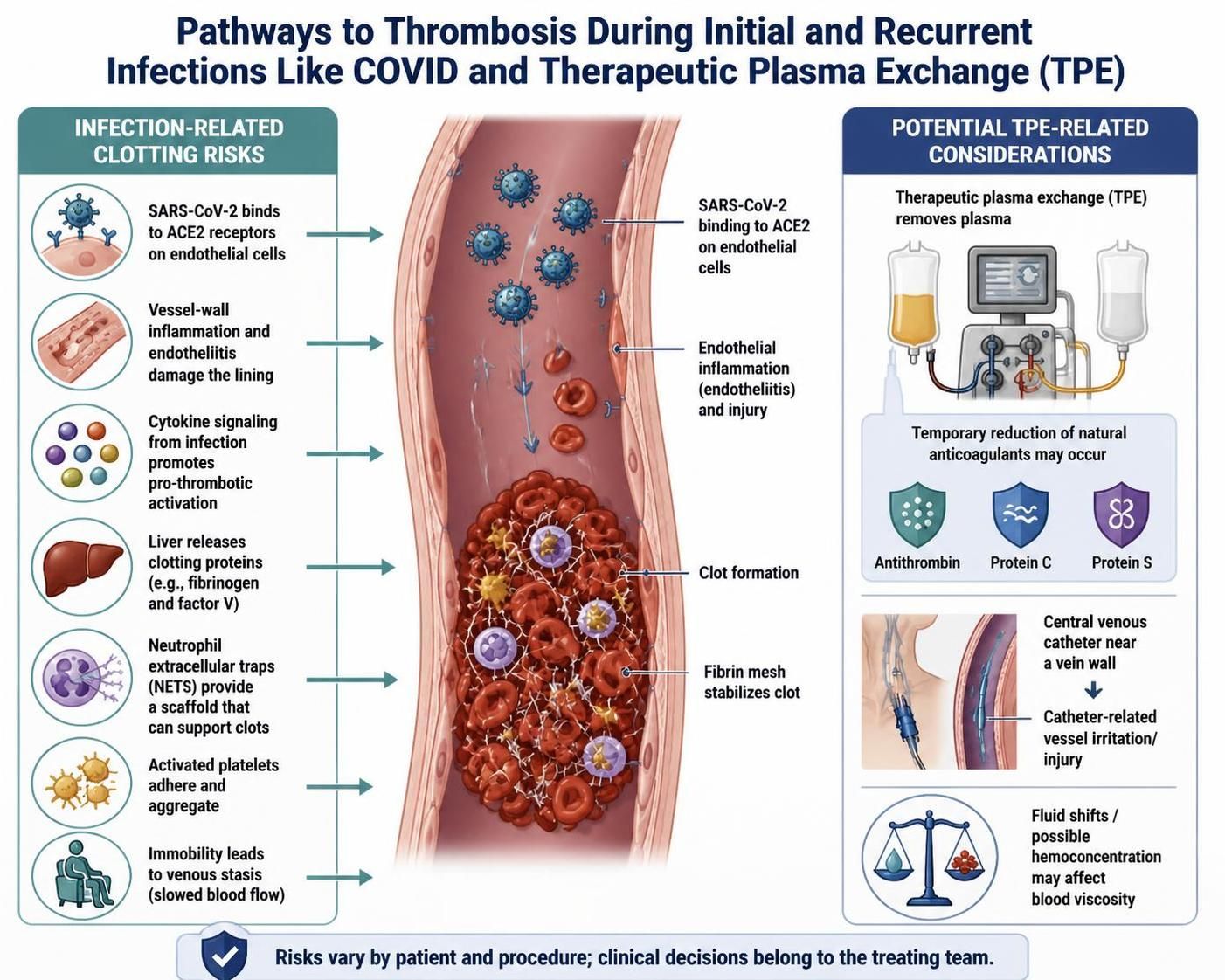
Recurrent deep venous thrombosis (DVT) during infectious diseases like COVID-19 happens because the virus triggers a severe systemic inflammatory and procoagulant state.
The mechanisms driving this increased risk are explained through Virchow’s triad (endothelial injury, hypercoagulability, and stasis) combined with specific immune responses:
1. Endothelial Injury and Viral Direct Invasion
-
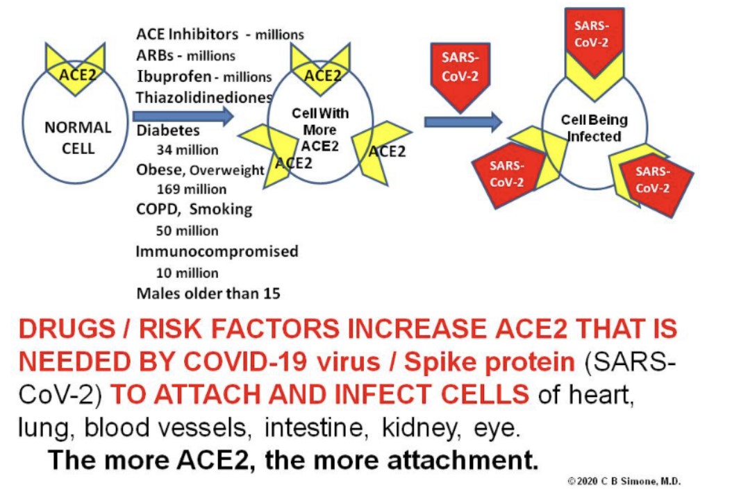
ACE 2 Receptor Binding: SARS-CoV-2 can directly infect and damage endothelial cells (the inner lining of blood vessels) by binding to ACE 2 receptors. The more ACE 2 receptors a person has, the more the attachment.
-
Viral Endotheliitis: This injury causes inflammation of the blood vessels, exposing underlying tissues that actively trigger the body’s clotting cascade.[1, 2]
2. Hypercoagulability (Overactive Clotting)
-
Cytokine Storm: A massive release of inflammatory cytokines during the infection causes the liver to overproduce clotting proteins—such as fibrinogen and factor V—at up to 2 to 3 times normal levels. [1, 2, 3]
-
Neutrophil Extracellular Traps (NETs): The immune system releases web-like structures called NETs to trap the virus, which inadvertently provide a scaffold for blood clots to form. [1, 2]
-
Platelet Dysfunction: Activated platelets clump together much more easily than normal. [1]
3. Venous Stasis (Slow Blood Flow)
-
Immobility: Bedrest, hospital or ICU admission, and general fatigue or weakness during a severe infection slow down blood circulation in the veins. Sluggish blood flow allows clotting factors to accumulate and form a thrombus. [1, 2, 3]
Why Prior DVT Patients Are at Higher Risk
-
If a patient has already had a DVT or has chronic vascular damage, their blood vessel walls are already altered or scarred.
-
When a systemic infection like COVID-19 hits, the sudden surge in inflammation and clotting proteins acts as a “second hit,” overwhelming natural anticoagulant mechanisms and precipitating a recurrent clot at the site of prior damage or in new vessels. [1, 2, 3, 4, 5]
Stopping an active clot and preventing a recurrent deep venous thrombosis (DVT) during an infectious disease like COVID-19 requires a combination of medical management to target hypercoagulability and physical strategies to counteract blood stasis. Because infectious diseases create a temporary but severe “high-risk window” that can last for weeks to months after infection, targeted interventions are critical. [1]
1. Stopping an Active Clot (Acute Management)
If a recurrent DVT occurs, the primary goal is to halt clot progression, prevent it from breaking off into a life-threatening pulmonary embolism (PE), and allow the body to naturally dissolve it. [1]
-
Parenteral Anticoagulants (In Hospital): For hospitalized or severely ill patients, doctors favor Low-Molecular-Weight Heparin (LMWH) injections (such as enoxaparin) over standard heparin. LMWH is more stable and does not interact negatively with certain antiviral medications like Paxlovid. [1, 2]
-
Direct Oral Anticoagulants (DOACs): For stable outpatients, physicians often utilize oral blood thinners like Rivaroxaban (Xarelto) or Apixaban (Eliquis). These medications target specific clotting proteins directly to halt the hypercoagulant cascade. [1, 2]
2. Preventing Clots During Infection (Prophylaxis)
Medical guidelines from organizations like the establish specific preventive protocols based on the severity of the illness:
-
Hospitalized Moderately Ill Patients: In-hospital patients who are not in the ICU but have high-risk markers (like significantly elevated D-dimer levels) may be placed on full therapeutic-intensity anticoagulation to aggressively get ahead of the systemic inflammation. [1, 2]
-
Hospitalized Critically Ill Patients: Counterintuitively, ICU patients are typically given standard prophylactic-intensity doses. Pushing to higher doses in critically ill settings does not show added benefit and sharply increases dangerous bleeding risks. [1, 2, 3]
-
Extended Outpatient Prophylaxis: While routine blood thinners are not recommended for everyone after discharge, patients with a prior history of DVT are considered high-risk. A doctor may extend a low-dose blood thinner for days or weeks after recovering from the virus to protect them during the post-infectious inflammatory window. [1, 2, 3, 4]
3. Physical and Lifestyle Defenses
To address the “venous stasis” (sluggish blood flow) caused by illness and bedrest, physical interventions must be deployed alongside medications:
-
Mechanical Compressions: Hospitalized patients who cannot take blood thinners due to bleeding risks rely on intermittent pneumatic compression (IPC) devices—sleeves attached to a pump that gently squeeze the calves to mimic walking.
-
Graduated Compression Stockings: For home recovery, doctors frequently prescribe prescription-grade compression stockings (typically 20–30 mmHg) to apply steady pressure, improve vein valve function, and accelerate blood return to the heart.
-
Early Ambulation and Calf Pumps: Even when isolated or fatigued, avoiding total sedentariness is vital. If confined to a bed or chair, performing ankle pumps (flexing the feet up and down repeatedly) activates the calf muscle pump, forcing blood out of the deep leg veins.
-
Aggressive Hydration: Fevers, sweating, and gastrointestinal symptoms from infections quickly cause dehydration, thickens the blood, and accelerates clotting. Drinking plenty of water keeps blood viscosity low. [1, 2, 3, 4, 5, 6, 7]
Summary of Interventions
| Goal | Medical Interventions | Physical & Lifestyle Interventions |
|---|---|---|
| Stopping an Active Clot | • Therapeutic-dose LMWH (inpatient) • Direct Oral Anticoagulants (DOACs) |
• Strict bedrest initially (per doctor instruction) • Elevation of the affected limb |
| Preventing a Clot (Inpatient) | • Prophylactic or therapeutic heparin based on acuity and bleeding risk | • Pneumatic compression boots • Frequent position changes |
| Preventing a Clot (Home/Recovery) | • Extended outpatient low-dose anticoagulants (for high-risk / prior DVT histories) | • 20–30 mmHg compression stockings • Regular ankle pump exercises • Active hydration (8–10 glasses of water/day) |
Yes, plasmapheresis can reactivate or trigger deep thrombophlebitis (deep vein thrombosis / DVT) through several specific mechanical and biochemical mechanisms. While the procedure is often used to remove harmful antibodies or inflammatory cytokines, it can temporarily create a highly prothrombotic (clot-promoting) environment. [1, 2, 3, 4]
The risk of reactivation or new clot formation during or immediately after plasmapheresis is driven by three main factors:
1. Depletion of Natural Anticoagulants
-
Albumin Replacement Fluid: When therapeutic plasma exchange (TPE) removes a patient’s plasma, it is commonly replaced with a 5% albumin solution. [1]
-
Loss of Protective Factors: Unlike fresh frozen plasma (FFP), albumin contains no natural blood thinners. The procedure strips away the body’s natural defenses against clotting—specifically Antithrombin III, Protein C, and Protein S—leaving the patient temporarily unprotected and highly prone to hypercoagulability. [1, 2]
2. Catheter-Induced Endothelial Injury
-
Central Lines: Plasmapheresis requires rapid blood flow, which usually necessitates a large-bore central venous catheter (such as in the jugular, subclavian, or femoral vein).
-
Vessel Trauma: The insertion and presence of this catheter cause direct physical trauma to the inner lining of the vein (endothelial injury). If a patient has a prior history of deep thrombophlebitis, this localized trauma combined with altered blood flow around the catheter can easily serve as the “second hit” that reactivates a clot.
3. Hemoconcentration and Fluid Shifts
-
Increased Blood Viscosity: The process of ultrafiltration and fluid shifts during apheresis can sometimes lead to mild hemoconcentration (thickening of the blood).
-
Clumping Risk: When blood is concentrated, the relative density of remaining procoagulant factors and platelets rises, slowing blood flow down and encouraging clotting cascade activation. [1]
Clinical Prevention Strategies
To prevent reactivation in high-risk patients, medical teams typically implement specific safeguards:
-
Prophylactic Anticoagulation: Patients with a history of DVT are often placed on a low-dose blood thinner (like low-molecular-weight heparin) before and during the course of apheresis therapy.
-
Partial Plasma Replacement: In some settings, physicians will mix fresh frozen plasma (FFP) into the replacement fluid rather than using 100% albumin to ensure that antithrombin III and other crucial clotting regulators are not entirely depleted. [1]
The combination of Myasthenia Gravis (MG), an active infection, and plasmapheresis creates a complex medical scenario where the risk of deep thrombophlebitis (DVT) must be tightly managed. Plasmapheresis—or Therapeutic Plasma Exchange (TPE)—is highly effective for clearing the pathogenic autoantibodies (like anti-AChR or anti-MuSK) causing an MG flare or crisis. However, balancing its benefits against the risk of recurrent clots requires highly specific medical coordination.
Why the Combination Raises Clotting Risks
-
Immobility from MG Weakness: Severe MG causes generalized muscle weakness, often affecting the legs, diaphragm, and bulbar muscles. This profound fatigue or partial paralysis leads to bedrest, which directly causes venous stasis (sluggish blood flow) in the lower extremities.
-
The “Triple Hit” Effect: A patient with a history of deep thrombophlebitis who is currently dealing with an infectious disease (like COVID-19) and undergoing plasmapheresis is experiencing three major clotting triggers at once: systemic viral inflammation, procedural depletion of natural blood thinners, and prolonged immobility.
Critical Considerations for Your Medical Team
Because of these overlapping risks, there are crucial adjustments the neuromuscular and apheresis teams typically make:
-
Heparin in the Apheresis Circuit: During the actual plasmapheresis procedure, an anticoagulant (usually ACD-A or standard heparin) is used to keep the machine lines from clotting. However, this only protects the blood while it is inside the machine, not necessarily the patient’s deep leg veins afterward.
-
Concomitant Outpatient Anticoagulation: The medical team must carefully balance therapeutic blood thinners (like Eliquis or LMWH injections) to protect the legs while ensuring they do not cause bleeding complications at the large-bore catheter insertion site.
-
Avoiding Certain Medications: If an infection or an MG flare causes respiratory distress, medications must be selected with extreme caution. For example, certain antibiotics or magnesium (sometimes considered for other issues) can dangerously worsen MG muscle weakness, making strict immobilization worse.
Structured Summary of Management Priorities
| Risk Factor | Impact on Myasthenia Gravis & DVT | Medical Management Strategy |
|---|---|---|
| Severe MG Muscle Weakness | Causes severe physical immobility, leading to sluggish leg blood flow (venous stasis). | • Immediate physical therapy for passive leg movements in bed. • Sequential compression devices (SCDs) on the calves. |
| Plasmapheresis (TPE) | Strips away natural anticoagulant proteins (Antithrombin III) during plasma removal. | • Monitoring of coagulation panels (PT/INR/PTT) post-procedure. • Consideration of partial FFP replacement if clotting markers spike. |
| Central Venous Catheter | Physical presence of the large line irritates the blood vessel wall, triggering local clots. | • Routine ultrasound tracking of the catheter site if swelling occurs. • Swift removal of the line as soon as the TPE cycle is complete. |